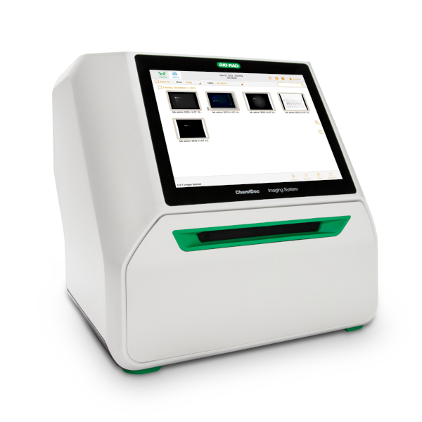
Bio-Rad ChemiDoc imaging and analysis system, designed for the acquisition, documentation, and analysis of images of gels and membranes in molecular biology and biochemistry applications. The equipment enables the visualization and recording of signals from applications such as gel electrophoresis, Western blotting, and other techniques using chemiluminescence or fluorescence detection.
The system combines a high-sensitivity CCD camera with an enclosed imaging chamber and illumination suitable for different detection methods, enabling the acquisition of digital images for subsequent analysis and quantification. The Image Lab software allows control of image acquisition, image optimization, densitometric analysis, and documentation of experimental results.
Calendar / Availability
Schedule Use
Implemented Methods:
Not Applicable
Possible Future Methods:
Not Applicable
Main Applications:
- Electrophoresis gel documentation;
- Western blotting;
- Detection by chemiluminescence;
- Protein and nucleic acid analysis;
- Densitometric band quantification;
- Signal intensity comparison between samples;
- Documenting and archiving experimental results;
Sample Types:
Main Advantages:
- High sensibility digital image acquisition;
- Refrigerated CCD camera for noise reduction;
- Closed system that reduces environmental light influence;
- Adequate for different gel and western blot documentation applications;
- Fast and reproducible image acquisition;
- Densiometric and quantitative analysis using Image Lab;
- Adjustable exposure and aquisition parameters;
- Documentation, analysis and data exportation tools;
- Simple user interface;
Technical Specifications:
| Specification |
Value |
| Manufacturer |
Bio-Rad Laboratories, Inc. |
| Model |
ChemiDoc |
| Software |
Image Lab Desktop |
| Detector |
CCD 6 MP, refrigerated |
| Dynamic Range |
>4 orders of magnitude |
| Smart Tray Technology |
Automatic recognition of application-specific tray and adjustment of imaging parameters and software options |
| Autofocus |
Precalibrated focus for any zoom setting or sample height |
| Auto-Exposure |
Two user-defined modes (rapid or optimal auto-exposure) for all blot and gel imaging applications |
| Image Flat Fielf |
Dynamic; precalibrated and optimized for each application |
| Maximum Image Area |
16.8 × 21 cm |
| Illumination Sources |
Trans-UV, 302 nm
Epi-white
Trans-white (requires White Sample Tray)
Trans-blue, 450-490 nm (requires Blue Sample Tray) |
| Detection |
Chemiluminescence filter
590/110 nm standard emission filter for protein
DNA gel and blot imaging
|
| Data Output |
16-bit ou 8-bit: SCN, TIFF, JPEG |
Documents: